The Anatomage table is a technologically advanced anatomy visualization system that allows users to explore the complex anatomy of the human body in digital form, eliminating the need for a human cadaver. The table presents a human figure at 1:1 scale, and utilizes data from the Visible Human effort with the additional capability of loading real patient data (CT, MRI, etc), making it a great resource for research, collaborative discovery, and the studying of surgical procedures. Funding to obtain the table was a collaborative effort between the schools of Dentistry, Movement Science, and Nursing although utilization is expected to expand to include Biology. Currently on display in the Duderstadt Center for exploration, the Anatomage table will be relocating to its more permanent home inside the Taubman Health Library in early July.
The Anatomage table allows users to explore the complex anatomy of the human body.
Technology Interventions for Health, $5M Center Award from Department of Education (UMHS, CoE, SI, Library)
Recently, the University of Michigan received a prestigious 5 million dollar Center Grant, awarded by the National Institute on Disability and Rehabilitation Research (NIDRR), part of the Department of Education.
The funds from this award will primarily be used to pursue several development, research, and training projects/studies involving technology interventions for self management of health behaviors. The newly formed center, led by Michelle Meade (PI, Rehab Medicine), will be an interdisciplinary endeavor, involving clinicians, researchers, and engineers from multiple departments on campus. This will allow UM researchers to continue to study how technology (including applications for smartphones/tablets, video games) can benefit individuals with spinal cord or neuro-developmental disabilities.
S.C.I Hard – an educational mobile game teaching independence to young adults with spinal cord injuries
For the past three years, the Duderstadt Center has been developing SCI Hard, a transformative game facilitating skill development and promoting the ability of individuals with Spinal Cord Injuries (SCI). Through game-play, SCI Hard teaches players how to manage their health and interact more readily in home, health care and community environments. Combining practical teaching methods with the element of play, SCI Hard aims to give autonomy and confidence back to individuals who find their world drastically altered after a spinal cord injury, specifically young men (ages 15-25) with a recent SCI.
Players navigate the game by wheelchair, enabling them to face their real-world challenges: juggling doctors’ appointments, attending therapy sessions to build muscle, and learning to drive a wheelchair-accessible vehicle. Even banal tasks such as waiting in line at the DMV are covered in a way that exposes the new obstacles individuals with a SCI may face. SCI Hard tackles this difficult subject matter with optimism and an earnest of humor. (The player’s quest is ultimately to stop the evil Dr. Schrync from taking over the world with zombie animals.)
Funds from this grant will be used to study how playing games like SCI Hard can directly benefit the health or alter the behaviors of individuals with a SCI, an effort that has been supported and well received by the accessibility advocacy, gamification, and health science communities. Receiving the Center Grant allows Duderstadt Center to continue to develop SCI Hard and other projects through Android support, more health/configuration options, voice acting throughout for greater immersion, and leader boards to help track progress.
To learn more about how the grant will be used and what University of Michigan departments are involved, read The Record’s write up on this great accomplishment. For a sneak-peek at SCI Hard and what it entails, check out the video below.
How can doctors and nurses walk around a hospital room that hasn’t been built yet? It may seem like an impossible riddle, but the Duderstadt Center is making it possible!
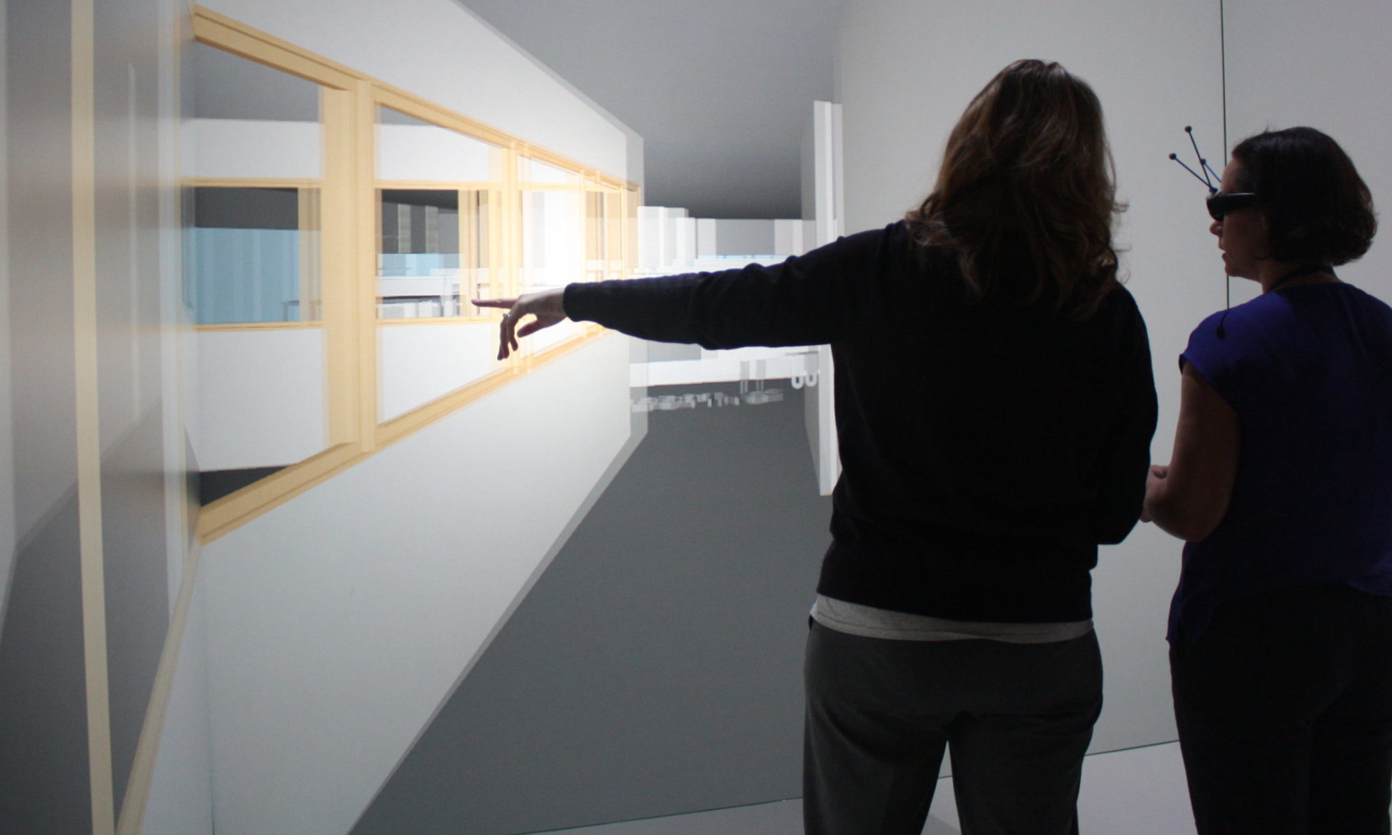
Working with the University Of Michigan Hospital and a team of architects, healthcare professionals are able to preview full-scale re-designs of hospital rooms using the MIDEN. The MIDEN— or Michigan Immersive Digital Experience Nexus— is our an advanced audio-visual system for virtual reality. It provides its users with the convincing illusion of being fully immersed in a computer-generated, three-dimensional world. This world is presented in life-size stereoscopic projections on four surfaces that together fill the visual field, as well as 4.1 surround sound with attenuation and Doppler Effect.
Architects and nursing staff are using the MIDEN to preview patient room upgrades in the Trauma Burn Unit of the University Hospital. Of particular interest is the placement of an adjustable wall-mounted workstation monitor and keyboard. The MIDEN offers full-scale immersive visualization of clearances and sight-lines for the workstation with respect to the walls, cabinets, and patient bed. The design is being revised based on these visualizations before any actual construction occurs, avoiding time-consuming and costly renovations later.
Ever have a headache or facial pain that seemingly comes and goes without warning? Ever been diagnosed with migraines, TMD or facial neuralgias but feel that your ability to explain your pain is limited?
PainTrek is a novel app that was developed to make it easier to track, analyze, and talk about pain. Using an innovative “paint your pain” interface, users can easily enter the intensity and area of pain by simply dragging over a 3D head model. Pain information can be entered as often as desired, can be viewed over time, and even analyzed to provide deeper understanding of your pain.
The PainTrek application measures pain area and progression using a unique and accurate anatomical 3D system. The head 3D model is based on a square grid system with vertical and horizontal coordinates using anatomical landmarks. Each quadrangle frames well-detailed craniofacial areas for real-time indication of precise pain location and intensity in a quantifiable method. This is combined with essential sensory and biopsychosocial questionnaires related to previous and ongoing treatments, and their rate of success/failure, integrating and displaying such information in an intuitive way.
Rebecca Davis, professor and researcher at the Grand Valley State University, received a research grant from the National Institute of Health to research how patients with Alzheimers disease navigate their living space. Assisted living homes can be drab or nondescript with long hallways adding to the confusion and frustration for those living in these homes. To research this problem and possible solutions, Davis recruited 40 people in the early stages of Alzheimer’s and 40 without the disease to virtually walk through a simulation of an actual assisted living home in the MIDEN. Staff and students at the Duderstadt Center modeled a 3D environment to re-create details such as the complicated lighting or maze-like hallways, to create a natural and immersive experience. This allows users to fully experience how the color schemes, lighting, and wall detail can affect the experience of living in the home. Various “visual cues” are placed throughout the space at key locations to determine if these help the subject in remembering which paths lead to where they need to go. Rebecca currently utilizes two environments in her study, one with visual cues and one without. Subjects are shown the path they must go to reach a destination and then given an opportunity to travel there themselves-if they can remember how.
University of Michigan News Services recently wrote an article on the release of PainTrek and it’s use for the treating pain in patients. PainTrek is an innovative computer/mobile application that measures pain area and progression in a particular subject or group of patients using a unique and accurate anatomical 3D system.
Through some innovative work done by Dr. Alexandre Dasilva and his team in the School of Dentistry, the Duderstadt Center was presented with some exciting new data related to migraines and their effect on the brain. We had to quickly turn the data into an image suitable for a pending journal submission. While we can’t go into details at this time about the research being done, we created a quick model of the data and brought it into the MIDEN for further exploration. The model was created by taking cross-sections of the MRI dataset and projecting those onto the surface of a brain mesh. The resulting model & textures were exported and then brought into the MIDEN.


 Through some innovative work done by Dr. Alexandre Dasilva and his team in the School of Dentistry, the Duderstadt Center was presented with some exciting new data related to migraines and their effect on the brain. We had to quickly turn the data into an image suitable for a pending journal submission. While we can’t go into details at this time about the research being done, we created a quick model of the data and brought it into the MIDEN for further exploration. The model was created by taking cross-sections of the MRI dataset and projecting those onto the surface of a brain mesh. The resulting model & textures were exported and then brought into the MIDEN.