Revolutionizing 3D Rotational Angiograms with Microsoft Hololens
A NEW WAY TO VISUALIZE THE HEART
Stephanie O’Malley
Just prior to release of the Microsoft Hololens 2, the Visualization Studio was approached by Dr. Arash Salavitabar in the U-M CS Mott Children’s Hospital with an innovative idea: to use XR to improve evaluation of patient scans stemming from 3D rotational angiography.
Rotational angiography is a medical imaging technique based on x-ray, that allows clinicians to acquire CT-like 3D volumes during hybrid surgery or during a catheter intervention. This technique is performed by injecting contrast into the pulmonary artery followed by rapidly rotating a cardiac C-arm. Clinicians are then able to view the resulting data on a computer monitor, manipulating images of the patient’s vasculature. This is used to evaluate how a procedure should move forward and to aid in communicating that with the patient’s family.
With augmented reality devices like the Hololens 2, new possibilities for displaying and manipulating patient data have emerged, along with the potential for collaborative interactions with patient data among clinicians.
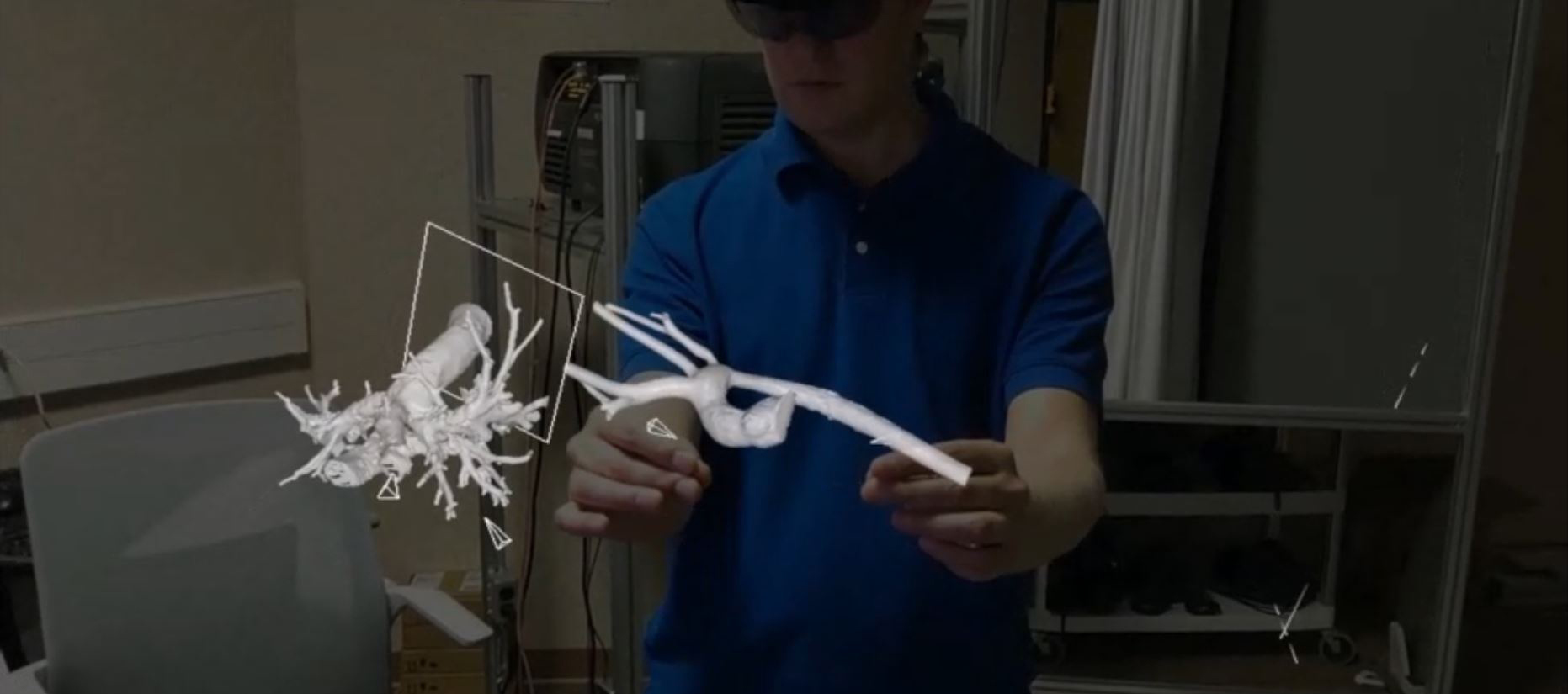
What if, instead of viewing a patient’s vasculature as a series of 2D images displayed on a computer monitor, you and your fellow doctors could view it more like a tangible 3D object placed on the table in front of you? What if you could share in the interaction with this 3D model — rotating and scaling the model, viewing cross sections, or taking measurements, to plan a procedure and explain it to the patient’s family?
This has now been made possible with a Faith’s Angels grant awarded to Dr. Salavitabar, intended to explore innovative ways of addressing congenital heart disease. The funding for this grant was generously provided by a family impacted by congenital heart disease, who unfortunately had lost a child to the disease at a very young age.
The Visualization Studio consulted with Dr. Salavitabar on essential features and priorities to realize his vision, using the latest version of the Visualization Studio’s Jugular software.
JUGULAR
The angiography system in the Mott clinic produces digital surface models of the vasculature in STL format.
That format is typically used for 3D printing, but the process of queuing and printing a physical 3D model often takes at least several hours or even days, and the model is ultimately physical waste that must be properly disposed of after its brief use.
Jugular offers the alternative of viewing a virtual 3D model in devices such as the Microsoft HoloLens, loaded from the same STL format, with a lead time under an hour. The time is determined mostly by the angiography software to produce the STL file. Once the file is ready, it takes only minutes to upload and view on a HoloLens. Jugular’s network module allows several HoloLens users to share a virtual scene over Wi-Fi. The HoloLens provides a “spatial anchor” capability that ties hologram locations to a physical space. Users can collaboratively view, walk around, and manipulate shared holograms relative to their shared physical space. The holograms can be moved, scaled, sliced, and marked using hand gestures and voice commands.
This innovation is not confined to medical purposes. Jugular is a general-purpose extended-reality program with applications in a broad range of fields. The developers analyze specific project requirements in terms of general XR capabilities. Project-specific requirements are usually met through easily-editable configuration files rather than “hard coding.”