Delving into the art (instead of science) of anatomy
New XR Course for FAll 2024
Author
Mary Clare Fischer
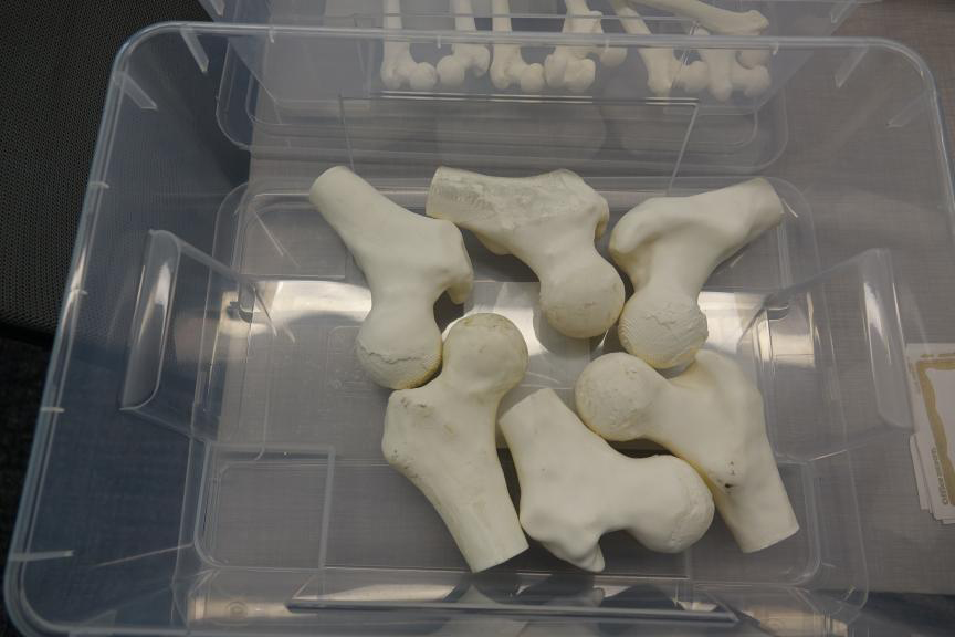
The first thing the students saw were the bones.
There were more than a hundred of them, stacked neatly in plastic bins on a long table in the front of classroom 2060. Some were long and slender, others bulbous and asymmetrical. All had the same glossy sheen.
From far away, they resembled delicate china figurines. Up close, it was easier to tell that they were 3D-printed versions of the same bones you’d find in a human pelvis or mouth or arm.
The students in the “Art of Anatomy” mini-course rummaged through the bone-like objects, serious expressions on their faces as they deliberated which to choose.
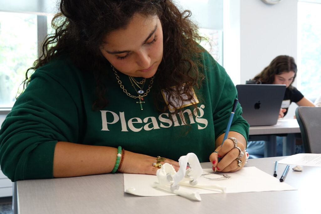
Their assignment that day was to create a sculptural arrangement. It could be anatomically correct; it could resemble nothing that you’d typically find in a skeleton. Then they had to take a picture of their designs and draw the shadows they’d made, using graphite pencils or charcoal.
The activity was intended to explore the body from a different perspective, to discover the angles and shapes its parts could make, and to ask the question that lay at the core of every session in this course: How does interacting with models of our anatomy, which try to approximate the experience of real human bodies, compare to encountering the real thing?
The next hour or so was nearly silent, except for the clunking of the tiny bones and the scratching of the pencils. Maya Moufawad, a pre-dental and art major, had chosen two halves of a jaw, complete with teeth. She fit them together and then affixed them to two smaller bones, making it look like the Flintstones had gotten their hands on a dental mold and decided to display it as art, using bones as the frame.
Movement science student Abby Kramer went with a thoracic vertebra, a lumbar vertebra, and a sacral bone. She liked being able to hold the bones, to turn them around and flip them upside down to better understand their structure and proportion.
She connected the lumbar vertebral bone directly with the sacrum, which would have been in the appropriate location anatomically. But, as she noted later: “There were still a lot of unknowns.” You couldn’t fully understand the body by looking at these bones. They were, both literally and figuratively, missing connective tissue.
“We’re trying to get them to understand that even the most factual anatomical model is still a fiction,” says Jennifer Gear, an art history and movement science lecturer who co-designed and taught the course. “It’s still removed from the body. In what ways and for what reasons? How do you stop thinking about these things as objective truths — but rather, to see them as believable fictions?”
***
When movement science student Regan Lee walked into the Capuchin Crypt in Rome, she, too, was fascinated by the bones.
In this case, they were real human bones from deceased Catholic friars, used to adorn a mausoleum that is like few others on Earth. The crypt is literally decorated with human remains — skulls framing archways, tibias and femurs arranged in elaborate crosses and mandalas on the walls and ceilings.
At the time, Lee was on a day trip during movement science associate professor Melissa Gross’ class, “Art and Anatomy in the Italian Renaissance,” for which students travel to Italy and use the classical statues and paintings of the Renaissance era as a guide to learning about anatomical structures.
As she walked away from the unique crypt, Lee was “nerding out.”
“I think everyone should see this,” she told Gross.
Gross had a different idea.
“What if we made this a class?” she pondered. “Let’s have students make their own art with the bones they’re used to looking at. We could 3-D print bones that the students could think critically about.”
“That’s crazy,” Lee responded. “Are you serious?”
***
Gross was indeed. She’d 3-D printed a small number of bones for previous anatomy courses, so she knew it could be done. And she’d spent her career creating innovative interdisciplinary courses in an attempt to engage students, stimulating them to learn in ways that worked better for them.
Together with Gear, she’d applied that paradigm of thinking to create the “Art and Anatomy in the Italian Renaissance” course Lee was so enjoying. She thought she and Gear could build off that successful partnership and come up with a class that challenged students to revisit their preconceptions about both art and the body.
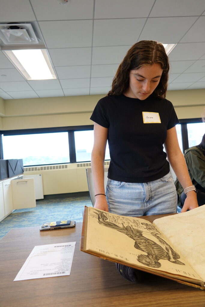
The pair started brainstorming. They wanted to teach a projects-based class — one with no tests, plenty of guest speakers, and lots of hands-on activities. They wanted to take students to different locations: the Hatcher Library’s Special Collections Research Center to look at Renaissance-era anatomy books, the Taubman Health Sciences Library to examine digital cadavers via the interactive Anatomage table, the Visualization Studio at the James and Anne Duderstadt Center on North Campus to play around with bones in virtual and augmented reality.
“I think of the classroom as a sandbox,” Gear says, “and I’m going to bring my best toys. Because I’ve got to be there with the students every day, too, and I don’t want to be bored. So I try to think about what would be fun to do, and this was a class that could lend itself to fun things.”
They wanted to ground the course in an arts-based approach, using critical thinking to respectfully challenge assumptions and foster dialogue that valued different perspectives. To do so, they planned to advertise in different schools on campus to attract students with varying backgrounds.
“Our goal was to open the students’ minds to other ways of seeing, of moving, of experiencing,” Gross says.
***
Coincidentally, the U-M Arts Initiative was looking for proposals for its Arts & Curriculum grant, which promotes the integration of arts into course development and teaching. In November 2022, the initiative gave its approval — and $19,611 worth of funds — to support Gross and Gear’s seven-week-long mini-course.
The pair used some of the grant money to pay Lee, who began the arduous task of printing the bones. Even the smallest ones took hours, and the printers often malfunctioned. Lee stuffed the ones that failed to print in her bag, and they clanked around as she walked.
“Even my apartment had bones everywhere,” Lee says.
Eventually, most of the bones made their way to SKB’s classroom 2060, as did 20 students — some from Kinesiology, some from Engineering, some from the Stamps School of Art and Design.
The students drew the bones, sometimes asking those who specialized in art to help the others portray the structures accurately.
They paged through 16th-century books full of woodcut illustrations of bodies and bones, their faces full of wonder at the opportunity.
They manipulated a digital cadaver on the Anatomage table, working as a group to make decisions about which bones and muscles and tissues to look at first and how to explore them. In that case, the Kines and biomedical engineering students often took the lead in explaining the names of the bones and where they were located.
They dissected five real animal carcasses and bones that Gross had gotten from generous butchers at Plum Market; one student, who disliked the smell of meat, was able to overcome her discomfort enough to participate with the support of her fellow classmates.
They talked about the ethics of using bones and bodies for research or education. In their reflection for that class session, students discussed whether they would donate their bodies to science given what they’d learned, noting that it was rare for them to feel this comfortable talking about such a difficult topic in class.
It began to feel like a kind of alchemy was taking place on Fridays from noon to 2 p.m.
“Every single class, I found myself being encouraged to think deeper, within my own knowledge and with the help of my peers,” one student wrote in a reflection. “The class’ emphasis on helping each other to understand is something I value so much. In fact, these discussions were so interesting to me that I always called my mom about them afterwards, because I was so excited to continue the conversation.”
“To see them bring their authentic selves to the challenges we’re setting every week, for them to treat it so seriously,” Gross says, “it feels like we’ve touched something important.”
***
On the final day of class, the students had one last opportunity to see the bones in a new way.
The Emerging Technologies Group at the Duderstadt Center had taken the digital files used to 3D print the bones and uploaded them to their visualization platforms, including virtual and augmented reality set-ups.
Movement science student Gordon Luo held a controller in one hand, using his index finger to press a button that grabbed the bone on his computer screen and moved it around. Then he found a way to digitally measure the bone.
“That’s so cool,” he says.
He was so immersed in the experience that he nearly tripped over the desk, less aware of his physical surroundings compared to the virtual world of the bones.
“It’s cool to realize this is where we’re at with technology,” he says.
Art student Summer Pengelly and biomedical engineering student Angel Rose Sajan were wearing HoloLens headsets that projected the bones hologram-style onto their surroundings.
“We’re building an elephant,” Pengelly tells me. “Or placing the bones so they’re shaped like an elephant head. I wish I could take a photo so I could show you. Oh, I just did.”
The photo was still contained in the software, so Pengelly picked up a piece of paper and started drawing the arrangement they’d made.
She and Sajan both agreed that they liked the HoloLens better than the VR headsets.
“It’s easier to manipulate the bones,” Pengelly says. “Using your hands as controllers gives you more access.”
“I kept turning the controller to figure out how to hold it,” Sajan says.
In the back of the visualization studio lay yet another digital environment to explore. Called the MIDEN for Michigan Immersive Digital Experience Nexus, it projects images onto the walls and floor of a room. Users wear headsets that place them within the environment created and give them tools to manipulate the objects in the space. In this case, students were able to slice a cadaver into different planes.
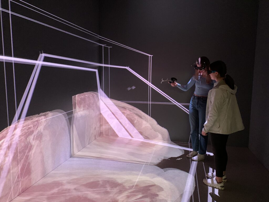
“MIDEN might be my favorite [of the technologies],” says Cece Crowther, a biomedical engineering student. “The Anatomage Table had the same energy as medical school. This felt more artistic.”
“But I could call three different [sessions] my favorite in this class,” she says. “Every class has been unique.”
***
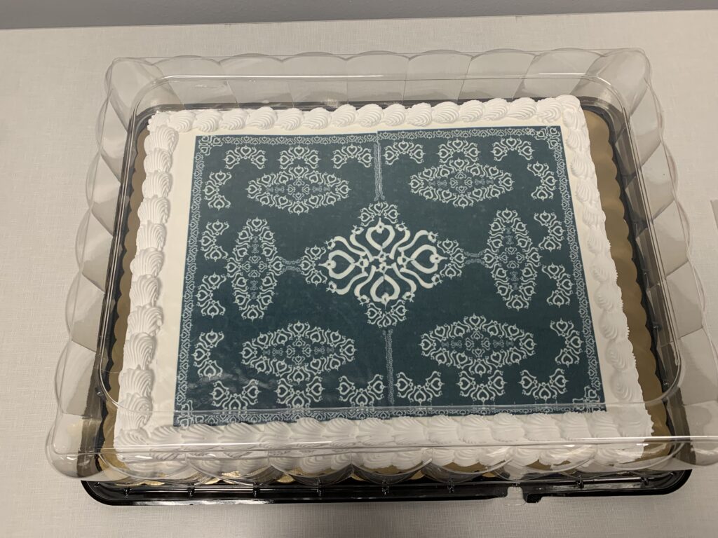
When the mixed reality class wound down, everyone gathered to eat celebratory cake. The top of the cake had an artistic design, made by creating a repeating pattern of one of the bone sculptures a student had designed early in the course.
“We’ve touched, looked at, manipulated, and drawn bones,” Gross says to the group. “Now we’re eating bones to wrap it all up.”
As students ate their cake, they reflected on the course, sharing feedback like, “I will not stop recommending this class to people” and “I made my schedule around this class.” Several mentioned that they’d gained so much from working alongside folks with different backgrounds.
“I appreciate this class so much because it normalizes the idea of art and science working together,” Moufawad, the art and pre-dental major, tells me. “Whenever I tell people what I’m studying, they always think it’s random, but it’s really not. There’s so much at the intersection of these two topics, and I love that this class celebrates that.”
A few weeks later, after the students have written their final reflections, I meet Gross in her first-floor office. She’s giddy over the success of the course. Her eyes light up and her tone becomes reverential as she talks about what she and Gear, with the help of some committed students, have managed to achieve.
“This experience we spent so many hours designing and thinking about, it actually worked,” Gross says. “Some important vein got exposed, and we’re not sure what’s flowing. It’ll take some time to unpack what was so empowering for so many students, but it’s a big fulfillment for us as teachers.”
“Delight,” she says, “is too soft a word.”
The Art of Anatomy course was made possible by a grant from the Arts Initiative at the University of Michigan to recipient Melissa Gross. Gross and Gear plan to offer the course again in fall 2024.
Full Article from the University of Michigan School of Kinesiology:
https://www.kines.umich.edu/news-events/news/delving-art-instead-science-anatomy