Learning Jaw Surgery with Virtual Reality
Jaw surgery can be complex and there are many factors that contribute to how a procedure is done. From routine corrective surgery to reconstructive surgery, the traditional means of teaching these scenarios has been unchanged for years. In an age populated with computers and the growing popularity of virtual reality, students still find themselves moving paper cut-outs of their patients around on a table top to explore different surgical methods.
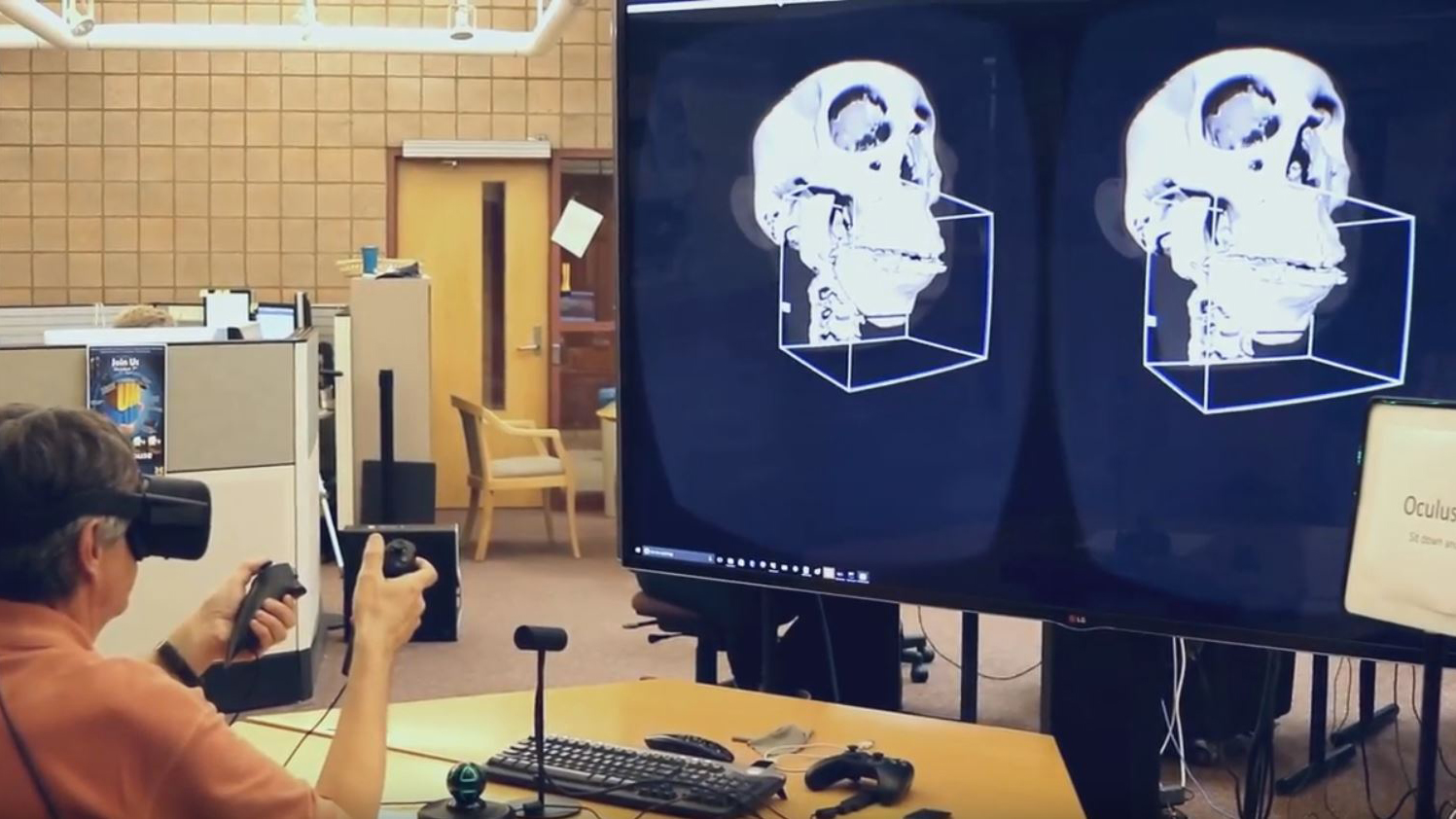
Dr. Hera Kim-Berman was inspired to change this. Working with the Duderstadt Center’s 3D artist and programmers, a more immersive and comprehensive learning experience was achieved. Hera was able to provide the Duderstadt Center with patient Dicom data. These data sets were originally comprised of a series of two-dimensional MRI images, which were converted into 3D models and then segmented just as they would be during a surgical procedure. These were then joined to a model of the patient’s skin, allowing the movement of the various bones to influence real-time changes to a person’s facial structure, now visible from any angle.
This was done for several common practice scenarios (such as correcting an extreme over or under bite, or a jaw misalignment) and then imported into the Oculus Rift, where hand tracking controls were developed to allow students to “grab” the bones for adjusting in 3D.


As a result, students are now able to gain a more thorough understanding of the spatial movement of bones and more complex scenarios, such as extensive reconstructive surgery, could be practiced well in advance of seeing a patient for a scheduled surgery.